
Presented at the 2012 SOFT Conference, St. Louis
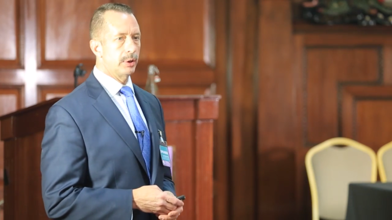
Dr. Carey began by saying questions were welcomed. He also noted most participants were familiar with the basics of trisomy, so he would emphasize new information. The result was he completed about 10% of his forty prepared slides. The order of information that follows reflects the questions that interrupted the prepared presentation.
John began by showing and explaining a karyotype, the diagnostic tool that dates back to the mid 1950’s. He explained that the reversed banding of the karyotype brings out the differences better. Chromosomes are paired and arranged by size, each pair numbered. Karyotypes are not as widely used as previously, with the advent of new techniques, which give more specific information and can measure specific changes in DNA. With karyotypes, cells are taken from blood lymphocytes, and a stimulant is given which makes the cells grow for two days, then additives are given to stop cell growth during metaphase, one of four stages of cell division. The cell is stopped just as it is about to split during mitosis. What is obtained is a picture of chromosomes stopped, but often curved, overlapped and not lined up. Previously, individual chromosomes were cut and pasted by students and physically lined up in pairs by size and counted. They were then photographed. The result was a karyotype. Many of us received this picture at our child’s diagnosis. Now a mouse pulls and drags the individual chromosomes and puts them in order, a much more efficient method.
There are more modern techniques to determine number and structure of chromosomes, in order to make a diagnosis. With the CGH microarray analysis, we now look at DNA and measure it to test for smaller, missing or extra pieces. The karyotype will never be a technique of the past, but it is not done as often.
An understanding of the structure of chromosomes is helpful. There is a centromere, the middle section or constriction, with an arm on either side. The smaller arm is the p arm, named for small in French, petite. The longer is the q arm, the next letter in the alphabet. This terminology came from a meeting in Paris in 1960, the same year that Patau and Smith in Wisconsin, and Edwards in England identified trisomy 13 and trisomy 18.
From the Human Genome Project we have learned about specific locations for specific disorders. We know the gene for cystic fibrosis is located at the top of the long arm of chromosome 7. BRCA 1, a gene for breast cancer is located on chromosome 17 near the neurofibrosis type 1 indicator. We know with adults with Down syndrome that memory and attention decrease in adulthood, and we now know the amyloid protein is on chromosome 21 and relates to memory loss and the development of Alzheimer’s disease. We have identified 18 genes on the 18th chromosome, now on a data base, but that does not help in understanding the findings of trisomy 18. We know colon cancer is on the long arm of 18 but It is not certain if this is related to the Wilm’s tumor prevalence in individuals with trisomy 18. There must be a gene on 18 that causes a heart defect, specifically a VSD, affecting the pumping chamber.
Of those with trisomy 18, 90% have a heart defect, usually a VSD, with polyvalvular heart disease, but more serious defects, truncus arteriosus, or hypoplastic left heart syndrome occurs in about 10% of those born with heart defects. The most common malformations must relate to a specific but still unidentified gene. It is not as clear what the effects of three copies of a chromosome are. Scientists use two mouse models for Down syndrome, actually creating a mouse with trisomy 16, the closest to Down syndrome. We need a T-18 mouse model. There is a macque monkey identified as having offspring with T-18, but primate labs are not as well tolerated by the public as rodent labs.
There is a specific technique for determining the extent of mosaicism. The protocol is to count 20 cells and look for mosaicism (some cells with an extra chromosome). This is the standard. This gives a 95% chance of picking up a cell with a different cell line. If one is found, then 50 or 100 more are counted. The number found then gives some idea of the percent of mosaicism: 15 of 100 cells with trisomy means 15% mosaicism. This was the best way of picking up cell lines of DNA before microarrays.
There is also the deletion syndrome, which means some genetic material is missing from a specific chromosome. This leads to manifestations more specifically than is the case with extra DNA. For instance, a missing segment on chromosome 13 puts one at risk for retinoblastoma. In contrast three copies of the same chromosome, thus the same segment, does not put one at risk.
Only some chromosomal trisomies are seen in live born infants. Chromosomes number 7, 8, 9 are seen in mosaic form in live born infants. In contrast, chromosome 11 and 14 in mosaic form are conceived but not live born. A few with mosaic 22 are live born, but rarely. There are fewer developmental genes in 13, 18, 21, so all three are live born with a full third chromosome. However, there is substantial fetal loss. Half of those identified with having trisomy 13 before 20 weeks are still born. Fetal loss is not so great with Down syndrome. The genes in chromosomes number 1, 2, 3 and 4 do not allow survival, which perhaps may be related to the placenta. Smaller chromosomes are generally more likely to result in a viable fetus, when there is an extra chromosome. There are exceptions which must relate to what genes are present. The 21st chromosome is bigger than the 19th, but 19 must include more developmental genes, because it cannot survive in a trisomy state. With chromosome 20, there are no live born, but it is a smaller chromosome than 21. The deciding factor in viability is not extra DNA but specific developmental genes.
There are more forms of aneuploidy (an abnormal chromosome count). Triploid, which means all chromosomes are in triplicate, are live born only if mosaic. This is the result of the breakdown of the natural mechanism that bars a second sperm from fertilizing an egg. There are 2 sperms, dispermia, each with a full complement of chromosomes.
It is believed that 60% of babies miscarried have a trisomy. One third are triploidy. Turner’s syndrome, XO in females, is a large cause of fetal death, but we are not sure why.
There is a range of human conditions involving chromosomal anomalies. There can be an alteration of number, which is an aneuploidy. This includes trisomy, monosomy, mosaicism of trisomy 18, 13 and 21, Turner’s syndrome (XO) and Kleinfelter’s syndrome (XXY).
There are also disabling and lethal conditions resulting from alteration of the structure of chromosomes. This includes deletions, duplications and translocations. An example would be 4p- in which a segment of the short arm of chromosome 4 is missing. A missing piece can be related to a rearrangement, translocation or reciprocal attachment, which affects chromosomal separation. With an unbalanced translocation a parent has the right amount of chromosomal material, but the child does not, getting too much of one chromosome and too little of another when the chromosomes aligned. Another way of dividing the chromosomes is into autosomal: 1-22, and sex chromosomes, X and Y.
Knowledge that has allowed us to identify specific chromosomes and understand the mechanism of specific syndromes has been recent. Only in 1956 was it possible to see the chromosome number. Only in the last 20 years have we been able to identify genes and place them on chromosomes. Progress in this area continues rapidly as more genes become known.
We also know statistics related to specific syndromes. We know 90% of children with trisomy 18 and 80% of children with trisomy 13 have heart defects. We know there is increased neonatal and infant mortality. We know 50% have succumbed by the second week of life. By six months of age, 50% have died. Only 10% will reach a first birthday. In the latest study from 2011 the numbers seem to stand the test of time. SOFT knows about the other 10%, the long time survivors.
The only way we know about survival is through data. A 1968 chromosome lab study says 3/4 of children who survive past a year, die by age 5. We have no new data confirming or denying this statistic. In Utah 10 children have lived past a year Ashton and one in Idaho, Erin in Utah. Three are still surviving, and seven, many known to SOFT members, have passed away at various ages over the past 20 years.
With trisomy 18 and 13 the ages of deaths spread out over next 18 years. Deaths of infants with trisomy 18 cluster in the first week, with more spread out in six months, then spread out over decades. The pattern holds. Doing more surgeries does not necessarily allow them to live longer. There can be maximum support, including cardiac surgery and immediate survival increases, but by a year survival rates remain the same. Trisomy 18 remains a highly serious condition.
There is data accumulated in Japan now, looking at heart surgery performed at the average age of 3 months. They went from 9% to 25% survival after heart surgery. Intervention did not seem to make much difference to survival. Children would pass away from something else such as pneumonia. There were 120 children and 89% had surgery and left the hospital.
All studies look at frequency of condition and there is the same live born frequency across cultures, and ethnicities. There has been 1: 5,000 to 1: 6,000 live born in Utah. With amnio and other prenatal diagnostic tools, the frequency numbers have changed, with live born dropping 40% as a result of elected terminations. There is greater availability and acceptability of termination. The updated data is in 2004-2009: 1 in 10,000.
Data in the United States from the Centers for Disease Control and Prevention (CDC) indicates there is a 70% termination of T-18/13, which is lower than the termination rate of Down syndrome which is 88-92%. In Europe, terminations are more frequent. Less common syndromes, such as T-9 mosaicism, which is rarer than T-13, have not had figures calculated. There has been a registry of chromosomal syndromes since 1999 in Utah, which will give more information. Not all birth defect registries keep track of all chromosomal syndromes.
We have much to learn of the outcome of survival. Procedures are increasingly available that can affect outcome. There are cardiac medications offered to some children, which can make a difference. For instance, cardiac meds are available which keep a PDA open, that is, keeps fetal blood circulation going which offsets some cardiac defects until surgery can be performed. When the PDA closes, which will happen naturally, the baby dies.
Time was over, many slides remained, but the session was informative, and participants, whose engagement was seen in questions and comments, wanted more time.


")


Recent Comments